Does your protein bind heparin — and which part of the sugar does it need? Heparan sulfate coats almost every animal cell, and hundreds of proteins use it: growth factors, chemokines, enzymes, and viruses looking for a way in.3 Heparin, its more highly sulfated relative, is inexpensive, well characterized and easy to attach to a sensor, which makes it the usual stand-in for heparan sulfate in binding studies.2,4 This guide shows how to measure protein–heparin binding with SPR — the surface, the controls, what the sensorgrams look like, and how a competition assay ranks heparin variants — using a 2020 study of the SARS-CoV-2 spike protein that was run on the P4SPR.1
Building a Heparin Surface
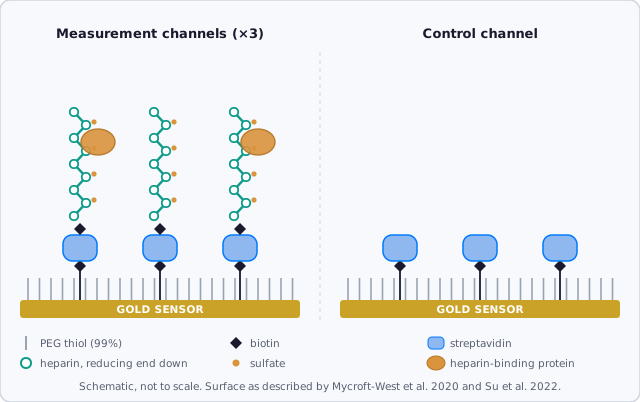
The surface decides whether you are measuring heparin binding or something else. Both studies cited here used the same design:1,2
- A PEG monolayer on gold, with 1% of the PEG chains carrying biotin. PEG resists protein adsorption, so the biotin sites are the only anchors.
- Streptavidin, injected over all four channels.
- Heparin biotinylated at its reducing end, injected over three channels. Attached by one end, each chain stands up from the surface much as heparan sulfate chains extend from their core proteins on a cell, leaving the whole chain free to bind.
- A control channel with streptavidin only. It tells you how much of your signal comes from the surface rather than from heparin.
The streptavidin step is the same as any biotin capture; our streptavidin capture protocol covers it on the P4SPR.
Check the Surface with a Known Binder
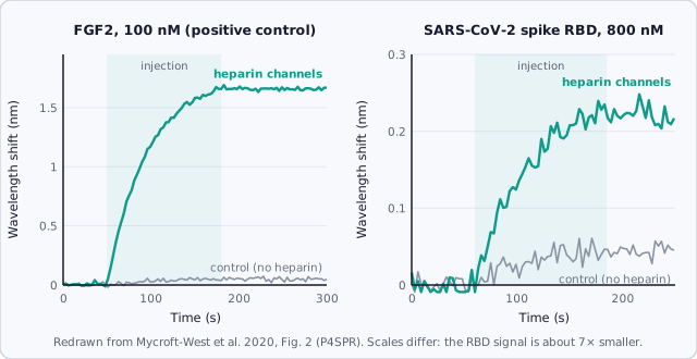
Before your protein, inject one whose heparin binding is well established. Fibroblast growth factor 2 (FGF2) is the usual choice. In the SARS-CoV-2 study, 100 nM FGF2 shifted the heparin channels by about 1.65 nm and left the control channel essentially flat; a 2 M NaCl wash removed it and restored the surface.1 Salt works because most heparin binding is electrostatic: positively charged lysines and arginines on the protein pair with the negatively charged sulfates on the sugar.3,4
The SARS-CoV-2 Spike: Binding That Doesn't Let Go
Injected at 800 nM, the receptor-binding domain (RBD) of the SARS-CoV-2 spike protein bound the heparin channels (about 0.22 nm), while the control channel showed about a tenth of that. The binding was to heparin, not to the layers underneath.1
Two things in these data are worth knowing before you run your own:
- It didn't dissociate. When the injection ended, the RBD signal stayed flat. A heparin surface is a dense forest of binding sites: a protein that lets go is caught by the next chain before it can leave. The authors therefore did not report kinetic constants — the data did not fit a one-site model — and noted that adding soluble heparin during dissociation is the usual way to stop this rebinding.1 Our sensorgram troubleshooting guide shows what rebinding and mass transport look like.
- Salt didn't clear it. The 2 M NaCl wash that strips FGF2 left the RBD in place. 20 mM HCl removed part of it; only 0.25% SDS removed it completely, followed by an HCl wash to clear the detergent.1 Scout regeneration from mild to harsh, as in our regeneration guide, and confirm your heparin still binds FGF2 afterwards.
Competition Assays: Ranking Heparins Without Kinetics
When dissociation can't be measured, competition still gives a clean comparison. Mix the protein with a soluble competitor, inject, and measure how much still binds the surface. Two details from the study keep the numbers honest:1
- Read the response after the injection, once the channel is back in running buffer. The bound RBD is still there, and any refractive-index difference between samples (competitors at mg/mL levels change it) has washed away.
- Inject the protein alone before every competitor and express each result as a percentage of that control, so slow changes in the surface don't bias the ranking.
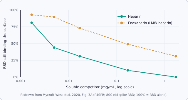
Heparin began to reduce RBD binding at 1.7 µg/mL and abolished it at 1.7 mg/mL. Enoxaparin, the low-molecular-weight heparin used clinically as an anticoagulant, also competed but was about 30-fold less potent by weight, and still left about 30% of binding at the highest dose.1
Which part of heparin matters
Chemically modified heparins, each missing specific sulfate groups, map what the protein recognizes. For the spike RBD:1
- Removing the 2-O- or 6-O-sulfates abolished competition; removing the N-sulfates only weakened it. The RBD prefers 2-O- and 6-O-sulfated regions.
- Fully desulfated heparin did nothing: the charges are essential.
- Over-sulfated heparin was the strongest modified competitor but still weaker than native heparin. More charge is not better if it stiffens the chain; the arrangement of the sulfates matters, not only their number.
- Short chains barely competed: an 8-sugar fragment had no effect and a 10-sugar fragment a modest one.
The same assay works well beyond viruses. The follow-up methods paper used it to show which sulfated structures a heparan sulfate sulfotransferase (an enzyme that adds sulfates to the chain) prefers, and that the drug suramin competes for its sugar-binding site.2
What Happened Next
The SPR work was one part of a larger study: in cell culture, heparin reduced SARS-CoV-2 infection of Vero cells by up to 80%, and circular dichroism showed that heparin changes the shape of the RBD.1 Later in 2020, another group showed that the spike binds heparan sulfate and ACE2 through the same domain, and that infection of cells depends on both.5 In the clinic, heparin was tested in COVID-19 mainly as an anticoagulant: in a large 2021 trial of hospitalized patients who were not critically ill, therapeutic-dose heparin improved survival to discharge without organ support compared with standard preventive doses.6 That trial was not designed to separate heparin's anticoagulant effect from any effect on viral binding.
Running It on the P4SPR
The four channels are what make this design practical: three heparin channels and a streptavidin-only control see every injection at the same time, so specificity is checked in the same run. In the 2020 study the P4SPR was run with an external pump. Today, binding checks and competition assays like these run with manual injection on the P4SPR 2.0, and the P4PRO with the AffiPump adds controlled flow when you need kinetics.
References
- C. J. Mycroft-West, D. Su, I. Pagani, T. R. Rudd, S. Elli, N. S. Gandhi et al., "Heparin inhibits cellular invasion by SARS-CoV-2: structural dependence of the interaction of the spike S1 receptor-binding domain with heparin," Thrombosis and Haemostasis 120 (2020) 1700–1715. doi:10.1055/s-0040-1721319
- D. Su, Y. Li, E. A. Yates, M. A. Skidmore, M. A. Lima and D. G. Fernig, "Analysis of protein–heparin interactions using a portable SPR instrument," PeerJ Analytical Chemistry 4 (2022) e15. doi:10.7717/peerj-achem.15
- D. Xu and J. D. Esko, "Demystifying heparan sulfate–protein interactions," Annual Review of Biochemistry 83 (2014) 129–157. doi:10.1146/annurev-biochem-060713-035314
- I. Capila and R. J. Linhardt, "Heparin–protein interactions," Angewandte Chemie International Edition 41 (2002) 390–412. doi:10.1002/1521-3773(20020201)41:3<390::AID-ANIE390>3.0.CO;2-B
- T. M. Clausen, D. R. Sandoval, C. B. Spliid, J. Pihl, H. R. Perrett, C. D. Painter et al., "SARS-CoV-2 infection depends on cellular heparan sulfate and ACE2," Cell 183 (2020) 1043–1057. doi:10.1016/j.cell.2020.09.033
- The ATTACC, ACTIV-4a and REMAP-CAP Investigators, "Therapeutic anticoagulation with heparin in noncritically ill patients with Covid-19," New England Journal of Medicine 385 (2021) 790–802. doi:10.1056/NEJMoa2105911