
IN SHORT
- What sticks: the coating you build on the gold decides what binds, how much, and what stays off.
- Two layers: first a thin linker on the gold, then the molecule you attach to it (the ligand).
- Choose from your ligand: pick the attachment that suits the molecule you have, then plan the experiment (TN-07: Designing a binding experiment). Regenerate with the mildest condition that brings the baseline back.
Overview
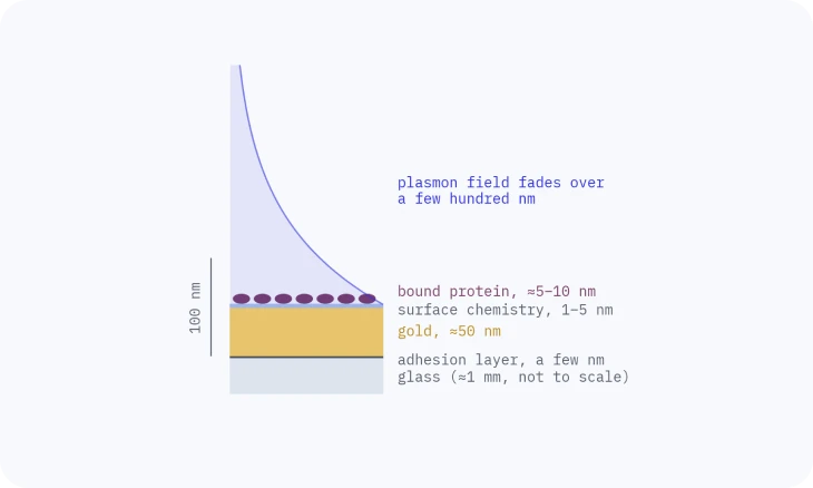
An SPR sensor only sees what binds within a few hundred nanometres of the gold (TN-01: What SPR measures). The binding surface decides what that is.
It has two parts: a linker layer on the gold, and the ligand you attach to it. Together they set three things: specificity (what binds), capacity (how much can bind) and fouling (what sticks that shouldn’t).
1 · THE LINKER LAYER
The linker layer
Gold does not hold proteins in a controlled way on its own. A thin organic layer goes on first, almost always through a gold–sulfur bond. How well that layer forms depends on the gold underneath: a smooth, clean film gives a dense, even monolayer (TN-05: The chip).
Alkanethiols pack side by side into a dense, ordered self-assembled monolayer (SAM), and the group at the top end of each molecule becomes the new surface.

| Linker | Top surface | What it is used for |
|---|---|---|
| MHDA (16-mercaptohexadecanoic acid) | Carboxyl (–COOH): hydrophilic, negatively charged at neutral pH | Covalent amine coupling of proteins and amine-modified DNA |
| AffiCoat | Peptide monolayer with carboxyl ends | Amine coupling with low background in crude samples |
| NTA | Nitrilotriacetic acid, loaded with Ni²⁺ | Capture of His-tagged proteins |
| Biotin + streptavidin | Streptavidin held on a biotinylated monolayer | Capture of biotinylated ligands, and the base for Protein A/G and other capture layers |
| Bare gold | Gold | Your own thiol chemistry: thiolated DNA, aptamers, custom SAMs |
2 · ATTACHING THE LIGAND
Three ways to attach the ligand
Covalent amine coupling (EDC/NHS). EDC/NHS turns the surface carboxyl groups into reactive esters, and lysines or the N-terminus of the ligand form a stable amide bond with them; leftover esters are blocked with ethanolamine. The bond lasts for many cycles. The cost: lysines sit all over a protein, so the ligand lands in random orientations, and some copies point their binding site at the surface.
Our standard recipe on MHDA: 30 mM EDC + 100 mM NHS (1:1), 4 min; ligand at 5 µg/mL in 10 mM sodium acetate pH 4.5, 30 s; 1 M ethanolamine pH 8.0, 6 min; a buffer rinse between steps.
His-tag on NTA. Ni²⁺ held by NTA grabs a His-tag, so every copy of the protein sits the same way up, with no activation step. The bond is reversible: imidazole competes with the His-tag and releases the protein; EDTA pulls the nickel out of the NTA, which releases the protein and empties the surface for recharging. The trade-offs: a slow drift as captured protein lets go, and no EDTA or imidazole in the running buffer.
Capture (streptavidin–biotin, Protein A/G, anti-tag antibodies). Streptavidin binds biotin so tightly that the bond is close to irreversible under normal assay conditions, so a biotinylated ligand stays put. Protein A or G on that streptavidin layer holds antibodies by their Fc tail, so the binding sites face the sample; low-pH glycine releases the antibody and the surface takes a fresh one.

| Covalent (EDC/NHS) | His-tag on NTA | Capture | |
|---|---|---|---|
| Orientation | Random | Defined | Defined |
| Holds the ligand | Permanently, for many cycles | Reversibly, with a slow drift as protein lets go | Biotin: stays put. Protein A/G: reversibly |
| Regeneration | Strips the analyte only | Strips the ligand too: reload each cycle | Protein A/G: fresh antibody each cycle |
| Our chips | MHDA (COOH), or AffiCoat for crude samples | NTA-His | Streptavidin, Biotin, Protein A |
| Choose it when you have | A stable protein with lysines | A His-tagged protein | A biotinylated ligand, or an antibody |
3 · ORIENTATION AND DENSITY
Orientation and density
How the ligand sits matters as much as how much of it there is. Oriented antibodies keep both binding sites free; randomly coupled ones lose a share of their activity.
More ligand is not always better: a crowded surface hinders binding, and a fast analyte meeting a dense surface can be limited by how quickly it arrives rather than by the binding itself (TN-08: Delivering the sample). For kinetics, aim for the lowest ligand level that still gives a clean signal.

4 · REGENERATION
Regeneration
Regeneration removes the analyte so the same surface can run the next cycle, without damaging the ligand. Start mild and step up only if the baseline does not return: for antibody–antigen pairs, glycine-HCl from pH 2.5 down to 1.5.
On capture surfaces you usually regenerate the capture layer instead (strip the antibody from Protein A, or the His-protein from NTA) and load fresh ligand.
5 · FOULING
Keeping everything else off
In serum, lysate or culture media, other molecules stick to any surface that lets them. Low-fouling layers reduce that background. Peptide monolayers from the founders’ academic group measured specific binding directly in crude cell lysate and in undiluted serum, and the same peptide platform supports His-tag capture. AffiCoat comes from that work. A reference channel and blocking handle the rest (TN-09: Getting a clean signal).
6 · CHOOSING A SURFACE
Choosing a surface
Start from the ligand you have. Try combinations in the Sensor builder, or ask us: tell us your molecules and your sample, and we will suggest a surface and a starting protocol.
| Your ligand | Start with |
|---|---|
| A protein with lysines, stable at pH 4–5.5 | MHDA (or AffiCoat in crude samples), amine coupling |
| A His-tagged protein | NTA-His |
| A biotinylated protein, peptide, DNA or glycan | Streptavidin |
| An antibody, and you want fresh ligand each cycle | Protein A or G |
| A thiolated oligo or aptamer, or your own chemistry | Bare gold |
Related
TECH NOTE · TN-05
The chip
What makes a good gold film, how films are made, and what degrades them.
TECH NOTE · TN-09
Getting a clean signal
Referencing, buffer matching, drift and complex samples: how to keep only the binding.
TECH NOTE · TN-10
From sensorgram to answer
Reading the curve, then affinity, kinetics and active concentration you can trust.