
IN SHORT
- The surface: molecules binding on the gold change the medium right at its surface (its refractive index). SPR only sees this thin layer.
- The dip in the light: the gold absorbs one colour of the reflected light, a dip. Binding moves the dip to a longer wavelength (why: TN-02: Why a gold film).
- The sensorgram: following the dip over time gives a real-time binding curve, with no label.
Overview
Surface plasmon resonance (SPR) follows molecules binding to a surface in real time, without labels.
It comes down to three steps: what binding does to the surface, how the light sees it, and how that becomes a binding curve. Knowing them tells you what a signal can and cannot tell you.
1 · THE SURFACE
What binding changes
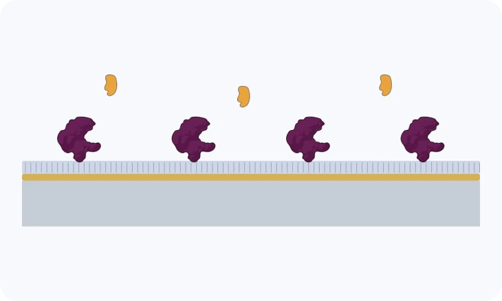
SPR watches a thin film of gold. When molecules bind to it, they replace water with denser material, and the medium right at the surface changes: its refractive index rises. That change is what SPR measures.
Only a thin layer counts. The sensor responds within about 200–300 nm of the gold and sees nothing beyond it (TN-02: Why a gold film). It does not detect a molecule's identity, and it does not weigh it directly.
For proteins and most biomolecules, the refractive index added is close to proportional to the mass added per area. That is why the response can be read as an amount bound, and why bigger molecules give bigger signals (Fig. 2).

2 · THE DIP
A dip in the reflected light
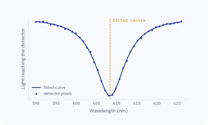
Light shone through a prism reflects under the gold film. At one colour, the gold absorbs it instead of reflecting it, and a dip appears in the reflected light. The light stays under the gold and never crosses the sample, which is why serum and lysate can be measured (TN-03: Lensless optics and wavelength readout).
When the refractive index at the surface rises, the dip moves to a longer wavelength (a red shift). The gold absorbs that colour because the light hands its energy to the electrons in the gold, a surface plasmon; TN-02 explains how, and why a gold film.
3 · THE SENSORGRAM
Following the dip over time
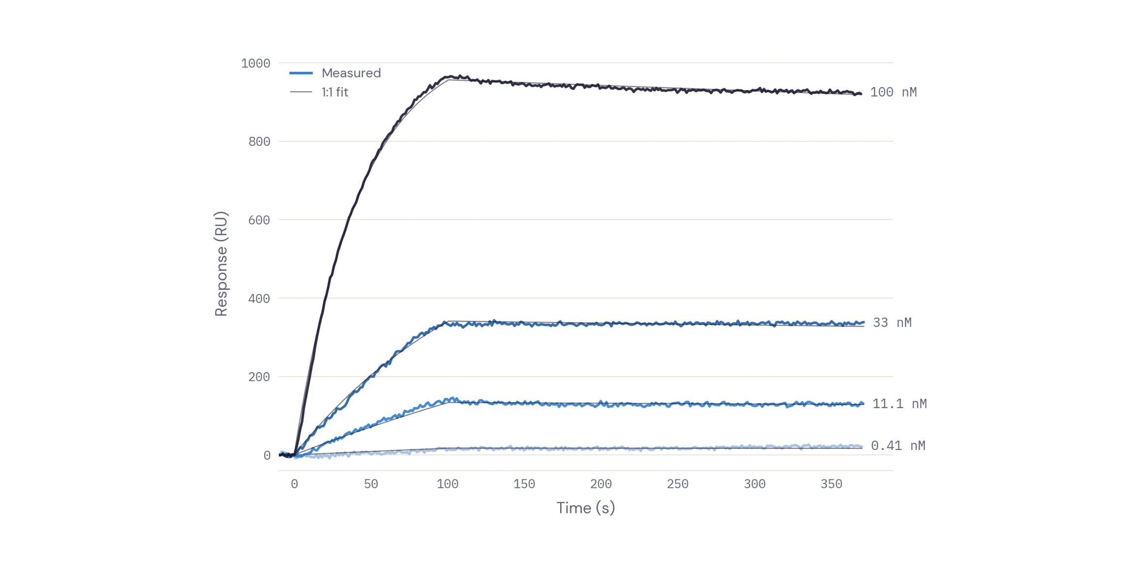
Following the dip position over time gives the sensorgram: a binding curve drawn as binding happens. It shows the association, the plateau and the dissociation, which is what lets SPR measure affinity and kinetics, not just presence (TN-10: From sensorgram to answer).
No fluorescent tag, no enzyme, no secondary reagent: the binding itself produces the signal.
4 · ITS LIMITS
What SPR can't tell on its own
Anything that changes the refractive index near the gold moves the signal. A sample buffer with a little more salt, glycerol or DMSO shifts the resonance without any binding; so do other proteins sticking where they shouldn't.
What bound is decided by the surface you build (TN-06: The binding surface) and the controls you run (TN-09: Getting a clean signal): a reference channel and blanks turn "the refractive index changed" into "my molecule bound".
Related
TECH NOTE · TN-04
Where the precision comes from
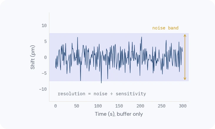
How the dip is located more finely than one pixel, and what sets the smallest shift you can see.
TECH NOTE · TN-06
The binding surface
Surface chemistries, coupling and capture, and how to choose one for your molecule.
TECH NOTE · TN-09
Getting a clean signal
Referencing, buffer matching, drift and complex samples: how to keep only the binding.